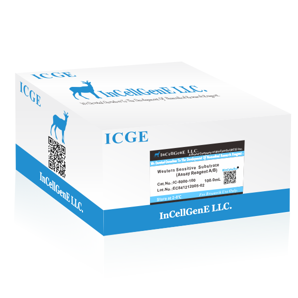
 DataSheet
IC-8000-500/500ml

Western Sensitive Substrate is an extremly sensitive ECL product by supplementing a proprietary luminol for chemiluminescent detection of immobilized proteins (Western blotting), conjugated with Horseradish Peroxidase (HRP) directly or indirectly. In Western blotting application, Western Sensitive Substrate provides high signal and low background, which allows detection of protein targets at femtogram levels. This feature benefits the researchers with excellent low background results and without signal burn effects at the same time.
Product Components
Western Sensitive Substrate (IC-8000-100)
|
Solution A (Luminol Solution) |
50 mL |
1 bottle |
|
Solution B (Peroxide Solution) User��s manual |
50 mL
|
1 bottle |
|
Western Sensitive Substrate (IC-8000-500) |
||
|
Solution A (Luminol Solution) |
50 mL |
5 bottle |
|
Solution B (Peroxide Solution) User��s manual |
50 mL |
5 bottle |
Please wear gloves, lab coat and goggles while operating. Prevent contact product directly. In case of contacting, wash with large amount of water.
Storage
Western Sensitive Substrate should be stored at 2-8 ��C and shielded from light. The product will be stable for 18months under 2-8��.
Materials needed but not provided
1.PVDF or nitrocellulose membrane
2.Wash buffer: Phosphate-buffered saline (PBS) or Tris-buffered saline (TBS) containing 0.05�C0.1% Tween®-20
PBS: 10 mM KH2PO4,150 mM NaCl, pH 7.4 TBS: 25 mM Tris, 150 mM NaCl, pH 7.4
3.Blocking buffer: 1�C5% (w/v) blocking agent (e.g., casein, BSA, or gelatin) in wash buffer
4.Specific primary antibody for interested protein, diluted in blocking buffer
5.HRP-conjugated secondary antibody, specific for primary antibody, diluted in blocking buffer
6.X-ray film or chemiluminescence image acquisition systems
Instruction
A.Protein transfer
1.Perform 1D or 2D electrophoresis for protein separation.
2.Move the electrophoretic gel into appropriate transfer buffer and equilibrate for 10 min- utes.
3.Wet the PVDF or nitrocellulose membrane in transfer buffer. (For PVDF membrane, it is necessary to pre-wet it in methanol before moving into transfer buffer).
4.Assemble the transferring sandwich as the order of two filter papers, gel, membrane and two filter papers.
5.Transfer proteins according to blotting apparatus manufacturer��s instruction.
B.Antibody incubation
1.Add BSA, skim milk based blocking buffer and incubate at room temperature for 30 minutes.
2.Prepare the primary antibody by diluting it with blocking buffer according to the manufac- turer��s instruction or previous experience.
3.Add primary antibody and incubate at room temperature for at least 1 hour with gentle agitation. For more specific interaction between primary and antigen proteins, it is recom- mended to perform additional incubation at 4 ��C for 8-12 hours.
B.Antibody incubation (~continued)
4.Decant the primary antibody solution thoroughly. Wash the membrane at least three times with ample amount of fresh Wash buffer for 10 minutes.
5.Prepare the secondary antibody by diluting it with blocking buffer according to the manu- facturer��s instruction.
6.Add secondary antibody and incubate at room temperature for 1 hour with gentle agita- tion.
7.Decant the secondary antibody solution thoroughly. Wash the membrane of at least four times with ample amount of fresh Wash buffer for 10 minutes.
C.Chemiluminescent detection
1.To prepare working HRP substrate, mix equal volume of Solution A and Solution B in a clean tube freshly. 0.1 mL of working HRP substrate is sufficient per 1 cm2 membrane area.
2.In the dark room or box, place the membrane side up in a clean box or plastic wrap. Add HRP working substrate onto the membrane.
3.Incubate the membrane at room temperature for 30 seconds.
4.Overlay plastic wrap or a transparency sheet on the wet membrane.
5.Expose the membrane to appropriate X-ray film or by chemiluminescence image acquisi- tion system. It is recommended to use 30 seconds as the initial exposure time.
D.Striping of PVDF membrane
1.Incubate membrane in stripping buffer (62.5 mM Tris-HCl pH 6.8, 100 mM ��-mercap- toethanol and 2% (w/v) SDS) for 30 minutes at 50-70 ��C.
2.Wash the membrane twice in Wash buffer for 10 minutes each.
3.To ensure complete removal of antibodies, incubate the membrane with HRP working substrate and expose against X-ray film for 5 minutes. No signal should be observed for complete stripping.
Order Information
|
Cat./REF. |
Size |
Price($�� |
Price(�) |
Price(��/CNY�� |
Price(��/JYP�� |
|
IC-8000 |
100ml |
$112.50 |
� 135.00 |
��1,125.00 |
��22,387.50 |
|
IC-8000 |
500ml |
$450.00 |
� 540.00 |
��4,500.00 |
��89,550.00 |
Troubleshooting
|
Problem |
Possible cause |
Remedy |
|
No signal or weak signal |
Poor transfer efficiency |
Optimize the membrane transferring procedure |
|
Insufficient antigen |
-Increase the amount of loaded antigen -Make sure the blot have been store correctly to avoid the degradation of target protein |
|
|
The concentration of primary and secondary antibody is too low |
Increase the concentration of the primary and/or the secondary antibody |
|
|
Inappropriate storage/preparation of the ECL detection reagents |
Use HRP or HRP conjugates to check the applicability of ECL reagents |
|
|
Too short exposure time |
Extend exposure time |
|
|
Excessive signal |
Antigen or antibody excess |
-Reduce the amount of loaded antigen -Dilute the primary antibody and/or the secondary antibody |
|
Antigen or antibody excess |
Optimize the condition by reducing the amount of antigen, or the concentration of the primary antibody and/or secondary antibody. Initially, reduce the secondary antibody to 20% of the original usage |
|
|
Inappropriate blocking |
Try different blocking substrate such as gelatin, casein, skim milk or casein |
|
|
High Background |
Inadequate washing |
-Increase the concentration of Tween-20 in washing solution -Increase the washing steps between the hybridization procedures -Extend washing time |
|
Overexposure to film |
Shorten the exposure time |
InCellGene LLC
A Brand Company Of BioCytoSci((BCS) INC.


